On my walks around Chicago, I pass dozens of COVID-19 testing sites drawing people inside with sandwich boards that read “PCR testing”. While PCR’s gold-star status in scientific research was well-established before the pandemic, it has since worked its way from scientific jargon into common vernacular.
The medical adaptation of PCR for COVID-19 testing illustrates the journey scientific research can take. Discoveries begin with a foundation in basic research— a type of research that aims to uncover the fundamental properties that make life possible. Once these basic facts are better understood, translational research uses those principles to develop novel technologies to improve human life. Furthering scientific innovation requires supporting basic research to continue broadening our knowledge of the natural world.
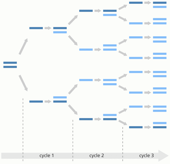
PCR, or polymerase chain reaction, is a primary tool in biological research that mimics the natural process of making copies of genetic material, but rather than occurring in a cell, it achieves this process in a test tube. Specifically, PCR amplifies relatively small pieces of DNA. It was a revolutionary invention for scientific research because it allows scientists to amplify small amounts of genetic material of interest to yield enough genetic material for a multitude of experiments.
COVID-19 testing determines if there is any of the SARS-CoV2 virus’ specific genetic material in a patient sample. The sample undergoes a process that converts the genetic material of the virus, RNA, into a corresponding DNA sequence. The sample then undergoes PCR testing, which recognizes if this viral DNA is present and makes enough copies to register positive on the test.
PCR uses heat to untangle the double-stranded DNA helix into a linear, single strand of nucleic acids. Nucleotides, the individual building blocks of DNA, come in four different types—cysteine (C), guanine (G), adenosine (A) and tyrosine (T). C and G bind together while A and T are partnered. During the PCR process, nucleic acids in the single strand bind with their complementary nucleic acid building blocks.
Left alone, nucleic acids would bind with their partners quite slowly. But introduce an enzyme, a biological catalyst, and the reactions pick up the pace. PCR uses a heat-resistant enzyme called Taq polymerase as a wingman to match up the nucleic acids along the single strand template with their binding partners, creating a new double strand of genetic material. The new double-stranded helix can again be separated with heat, creating even more strands for the enzyme to pair up. When the original sequence’s complementary strand is matched up with its partnering nucleic acids, the original strand is regenerated. This cycle is continued numerous times, copying one strand into millions.

Taq polymerase, the workhorse of PCR, was detected in an unlikely place over 50 years ago. This enzyme was discovered in 1969 by a research team studying bacteria living in the bubbling hot geysers in Yellowstone National Park. At high temperatures, the enzymes that enables the biological reactions of life can become inactive, like how frying an egg deactivates the proteins in egg whites, turning them from clear to white. The researchers discovered that these heat-loving bacteria had specialized enzymes that were active even at high temperatures, making them compatible in processes that require heat, like untangling DNA.
In 1983, Dr. Kary Mullis invented modern PCR by combining the background knowledge of known properties of DNA with the discovery of the heat-resistant Taq polymerase enzyme at Yellowstone. Almost 10 years later, Mullis was awarded the Nobel Prize in Chemistry for his invention. The Yellowstone researchers likely never imagined that poking around geysers would lead to such a revolutionary technology. The team’s quest for basic understanding of how life was possible at high temperatures paved the way for Mullis’ translational application.

To fuel ongoing translational research, we must continue to expand our foundational knowledge. As a Ph.D. student at Northwestern University’s Driskill Biomedical Graduate Program, I conduct basic research determining how epithelial tissue grows and develops. The epithelium is a thin sheet of cells that lines organs, acting as a barrier and compartmentalizing the body. The skin is the most famous example of epithelium tissue, engulfing the entire body and protecting it from outside elements.
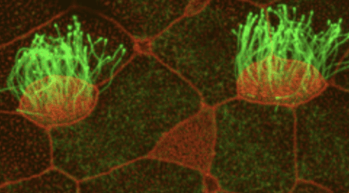
I use developing frog embryo skin to study epithelial growth. Frog embryo skin is like the epithelium that lines the lungs and fallopian tubes in that it also contains multiciliated cells (MCCs). MCCs are cells that have over 100 hair-like appendages called cilia emanating from the surface, like a surrealist sea anemone. In the lungs, cilia push down mucus to protect our respiratory organs; in the fallopian tubes, cilia guide egg cells down the tubes.

MCCs embed into the epithelium through a process called radial intercalation. MCCs begin to develop below the surface and squeeze upward between neighboring cells to join the outer epithelium, like Destiny’s Child’s Kelly and Michelle rising from beneath the stage to join Beyoncé at the Superbowl.
To enter the epithelium, MCCs must bypass an obstacle course of tightly bound junction proteins that are responsible for making the epithelium a barrier. My research goal is to understand the communication between MCCs and their neighboring cells that allow MCCs access into the epithelium.
My research program is associated with Northwestern University’s Feinberg School of Medicine because of its translational applications in the medical field. While it may seem a stretch to connect studying embryonic frog skin to preventing cancer metastasis or developing synthetic organs, it’s not far off. Cancer cells also radially intercalate between cells to enter the bloodstream and spread throughout the body. Similarly, understanding how junction proteins respond to tension in the epithelium could help scientists shape flat sheets of epithelial cells into 3D, lab-grown mini organs for future organ transplantations.
Basic research might appear to be science for science’s sake. However, the wealth of knowledge that can be gained from such research has the potential to spur revolutionary breakthroughs that vastly improve our lives. Sometimes, a journey that begins with probing a hot geyser for biological life can end in PCR-based COVID-19 testing sites throughout Chicago.
The post From Geysers to COVID Testing: The Crucial Contributions of Basic Research appeared first on Illinois Science Council.


Leave a Comment